Key features :
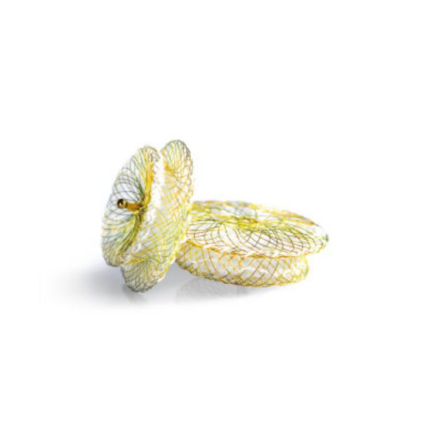
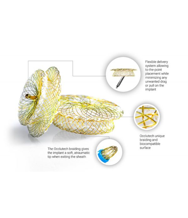
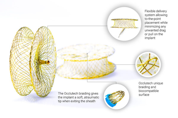
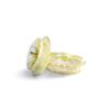
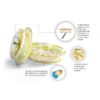
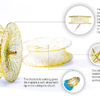
- Unique Braiding Technology: No distal hub protrusion, offering flexibility and adaptability.
- Flexible Design: Reduces the risk of damage to surrounding anatomy.
- Ball-and-Socket Design: Allows a wide range of angulation between the device and the delivery cable.
- Symmetrical Device Design: Ensures proper alignment and complete closure of the defect.
Sizing and Compatibility
Occlutech mVSD Occluders are available in various sizes to accommodate different defect dimensions:
- Transesophageal Echo (TEE) Sizing: Utilizes specific angles and views for accurate measurement.
- Angiography Sizing: Involves contrast injection via a catheter in the left ventricle to measure the VSD. The occluder should be sized based on the actual defect size or 1-2 mm larger than the maximum VSD size at the end of diastole.
Product Specifications
Occlutech provides a detailed compatibility chart for different mVSD occluder sizes, ensuring optimal fit and performance.
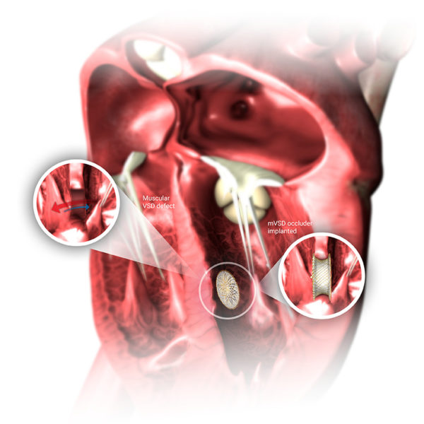
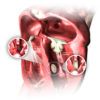
A Ventricular Septal Defect (VSD) is a congenital heart defect characterized by a hole in the septum that separates the heart’s two lower chambers (ventricles). Isolated VSD accounts for 37% of all congenital heart diseases in children. The incidence of isolated VSD is about 0.3% in newborns, as up to 90% may eventually close spontaneously. The incidence is significantly lower in adults. VSDs have no gender predilection.
Symptoms of VSD
Symptoms can appear in newborns or later in life. While many VSDs close spontaneously, significant defects can lead to serious complications such as:
- Pulmonary Hypertension (PAH)
- Ventricular Dysfunction
- Increased Risk of Arrhythmias
Types of VSD
VSDs vary based on their morphology:
- Type 1 (Infundibular Outlet): Located beneath the aortic and pulmonary valves in the right ventricular outflow septum, accounting for only 6% of all VSDs.
- Type 2 (Membranous): The most common type, representing 80% of all defects. If it involves the muscular septum, it is called perimembranous.
- Type 3 (Inlet or Atrioventricular Canal): Located just below the tricuspid and mitral valves in the right ventricular inlet septum, representing 8% of all defects. Commonly seen in patients with Down syndrome.
- Type 4 (Muscular Trabecular): Found in the muscular septum, typically in the apical, central, and outlet parts of the interventricular septum. Represent up to 20% of VSDs in infants but are less common in adults due to the tendency for spontaneous closure.
Types 2 and 4 are suitable for transcatheter VSD closure.

